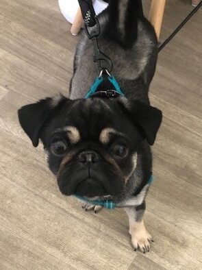
Frankie, a sweet little pug was referred to our neurology team in October last year when he was only 17 weeks old. His owner was concerned as over the previous 5 days he had progressively lost the ability to stand or walk and appeared to be in pain.
After a number of tests including an MRI scan, an abscess was found on his spine. Discussing the options and due to his age and size it was decided that Frankie would be given some medication and antibiotics and his owner would watch to see if this made an improvement. When Frankie came back a month later it appeared he had responded very well to this medical management, and he was sent home happy with a notes that if it was a foreign body, he may need more scans and surgical intervention.
Frankie was re-checked in late November and was recovering remarkably well, more playful and brighter and with a gradual improvement of the neurological function of his back legs. Things were looking good, then at the start of December he was re-admitted as the function in his back legs had suddenly deteriorated. He had an MRI and Ultrasound scan and that revealed an infection and the abscess again as a migrating foreign body causing compression of his spinal cord and therefore the paralysis. It was again decided to go down the conservative rather than surgical route but to keep an eye on Frankie to see how comfortable he is.
Unfortunately, Frankie had to be referred to us again as he was in pain and showed signs of deteriorating neurological function. He was referred to our Soft Tissue Surgery team was sent for a CT scan. This scan showed an abnormal tract and soft tissue swelling from his diaphragm to beneath his sublumbar muscles – seen by the pink arrows.
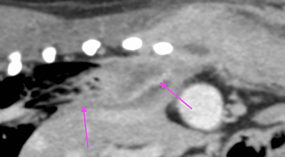
There was also a collapse of the disc space between his lumbar verterbrae 1-2 and erosion of his verterbral bones – the green arrow.
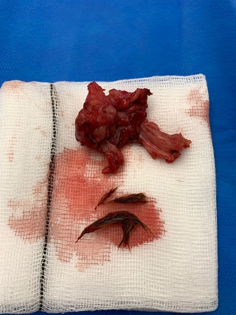
Because of the pain Frankie was in and these dramatic changes, the team decided to take Frankie to surgery after discussing the risks involved with his owners that included permanent paralysis. Frankie had a complicated surgical procedure as Jackie one of our Soft Tissue Consultants had to open up both his thoracic and abdominal cavities and follow the soft tissue tract from his diaphragm to lumbar vertebrae. During this process they had to remove part of his lung lobe as this was attached to his diaphragm. They entered the spinal canal and found an extensive abscess and retrieved two foreign bodies (grass awns) then flushed the area and took samples for the pathology team to investigate and then closed his thoracic and abdominal cavities.
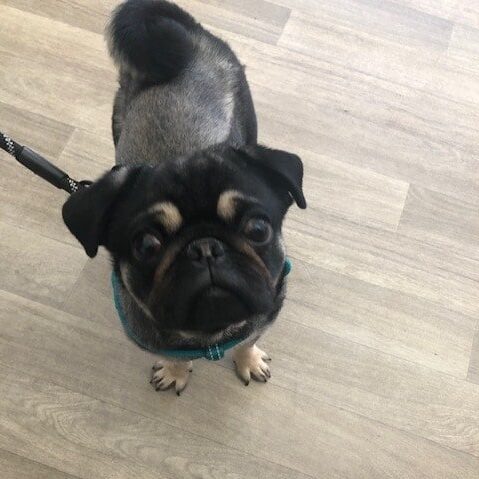
After a great deal of nursing care of his wounds with the nursing team, a chest drain and the physiotherapy team creating a rehabilitation programme for him, Frankie was eventually discharged. The team saw him back a month after and he was like a different dog, bright, active and with no evidence of pain at all! His neurological exam showed significant improvement of his ataxia (loss of co-ordination). His owners were absolutely delighted!
It is very likely that Frankie had inhaled the grass awns and they had travelled from his mouth or nose and tracked through his lungs, diaphragm and sublumbar muscles. Without surgery to remove the grass awns he would not have improved. Grass awns are very difficult to identify on any scan not only because they are small but the location they managed to get to location. Thankfully the brilliant team found them with the intricate surgery and Frankie went on to make an excellent and full recovery.