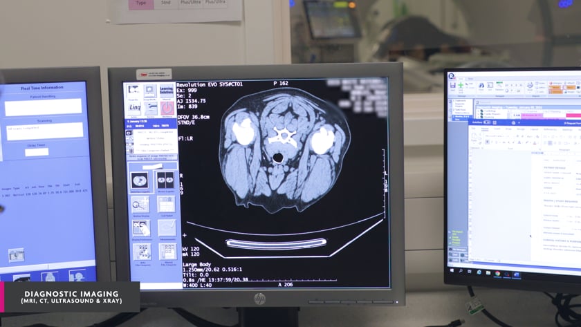
Diagnostic Imaging is the creation of visual representations of the inside of a body.
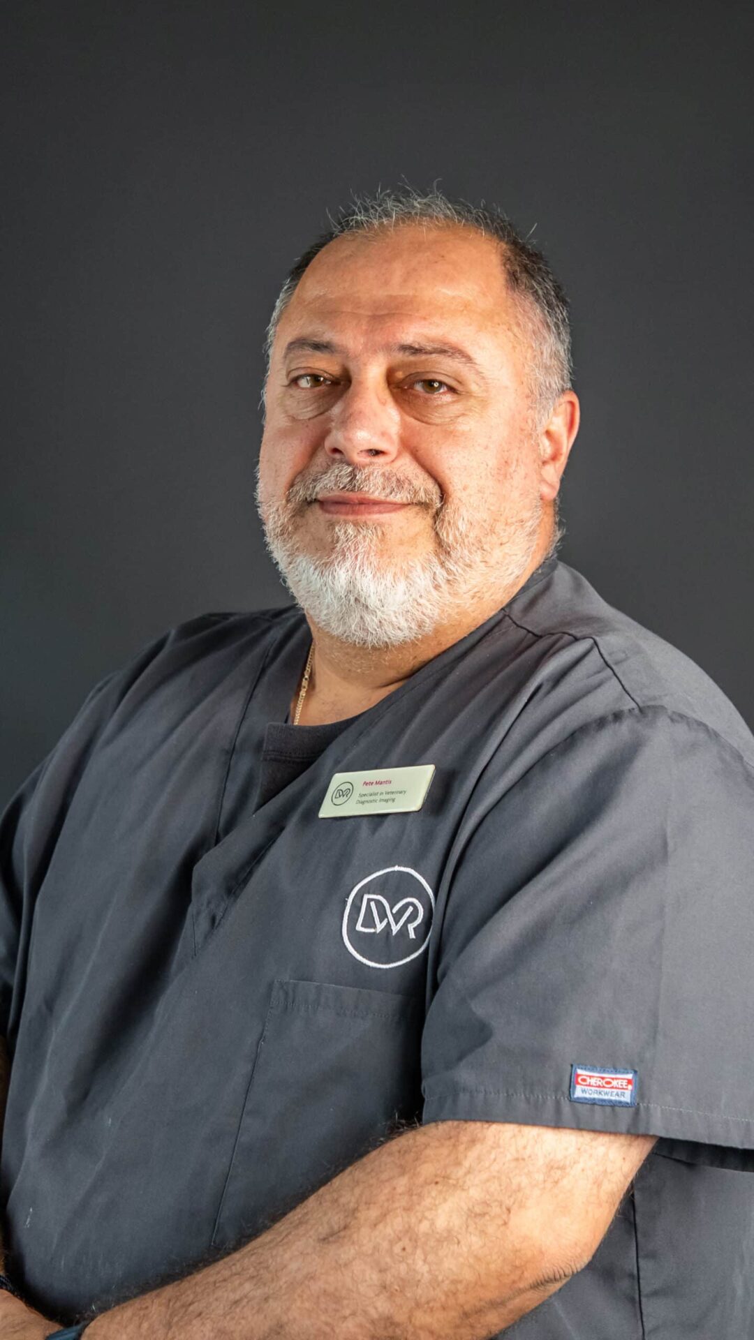
Led by Pete Mantis, this essential service supports all the clinical areas by providing images of internal structures such as muscles, tendons, bones, nerves and internal organs. These images are used to diagnose and facilitate treatment in our patients, greatly contributing to the success of the outcome.
We are also able to perform minimally invasive procedures under imaging guidance, such as fine needle aspirations or biopsies of diseased organs and removal of foreign material under ultrasound guidance. This means that diagnoses can be reached and treatment facilitated without subjecting the animals to more invasive surgery.
Digital radiography is a form of imaging in which sensors are used instead of traditional photographic film. The x-ray images are available instantly, without waiting for processing, and they can immediately be shared with colleagues at their desk, consulting room or operating theatre throughout the clinic.
Ultrasound is a safe and painless procedure, which allows sound waves to be reflected by internal body structures, creating ‘echoes’ that are converted into live images. This technique is used to visualise muscles, tendons, eyes, blood vessels and many internal abdominal or thoracic organs.
Our MRI scanner provides high quality images with excellent soft tissue differentiation, without the use of radiation. MRI scans are particularly useful in the diagnosis of neurological diseases, but also in some orthopaedic conditions, soft tissue trauma, foreign bodies and tumours.
CT scans can be preferable when looking at parts of the body such as lungs, the nose, joints, bones and abnormal blood vessels. The scanner acquires very thin x-ray images which are converted to 3-D by a computer. CT angiography allows more detailed evaluation of the blood vessels for identifying abnormalities and for planning corrective surgery.